Imaging tells the story the stethoscope can’t. Clear views. Clean measurements. Calm guidance when a patient is worried. Diagnostic Cardiovascular Sonography builds those habits. You learn to capture standard views on demand, speak in plain language, and hand a cardiologist data they can act on. Technical, yes. Also very human.
What Diagnostic Cardiovascular Sonography Looks Like Day One
Start simple. Patient ID. ECG leads. Machine presets. Then probe grip, wrist angle, and pressure you can hold for sixty seconds without shaking. You run through the parasternal long axis, rotate to short axis, slide to apical four, and finish subcostal with the IVC. Diagnostic Cardiovascular Sonography turns that sequence into muscle memory. Set depth, trim gain, freeze, label, save. Repeat until it feels boring. Boring is good. It means consistent.
Physics You Actually Use
You do not need to love equations to use physics well. Frequency sets resolution. Depth trades detail for reach. Artifact has a cause, so you fix it on screen instead of fighting the patient’s position. In the lab, you tweak TGC, narrow color boxes, and learn when to move your body rather than your wrist. Small changes, big results.
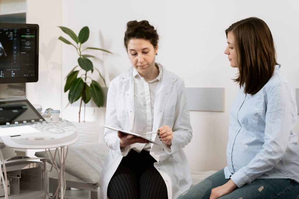
Patient Communication That Lowers the Heart Rate
Many patients arrive short of breath or afraid of what the scan might find. Scripts help, but tone matters more. One sentence upfront: what you will do and how long it takes. A heads up before pressure on the rib space. Check comfort when you roll them into left lateral decubitus. Offer a blanket. Keep directions short. One step at a time. The exam goes faster when the patient trusts you.
Quality and Safety You Practice Every Shift
Clean machine. Fresh gel. Disinfect between exams. Confirm the name on the screen before the first image. For contrast or stress environments, follow the site protocol and stay inside your scope. A short checklist prevents repeats. It also protects your patient and your lab.
Clinical Rotations: Where Rhythm Sets In
Class builds knowledge. The clinic builds timing. You set rooms, pace add-ons, and keep the schedule honest without rushing a difficult exam. You will scan real cases: valve disease, wall motion changes, pericardial effusions, post-procedure checks. What preceptors want early is steadiness. Complete views. Measurements in the right order. Worksheets that read cleanly. Speed arrives after you get the order and the hand control right.
Tools You’ll See and Use
Cart and portable systems. ECG integration. PACS for storage and review. You will route studies, verify they reached the queue, and handle small troubleshooting without drama. Learn the few settings that matter most at each site. Also learn how to reset safely when something looks wrong.
Where Diagnostic Cardiovascular Sonography Leads
Graduates step into hospital echo labs, outpatient centers, and cardiology practices. Early work centers on transthoracic exams. With site training, some support stress testing or assist around transesophageal workflows. Over time, you mentor new students, help refine protocols, or add vascular coverage. The core stays the same. Reliable images. Clean numbers. Patient care that feels steady.
Closing Section
Diagnostic Cardiovascular Sonography blends precision with bedside calm. You build repeatable views, collect trusted measurements, and guide patients through a technical exam without turning it into a maze. If you want the specifics on coursework, labs, and rotation flow, start with the Diagnostic Cardiovascular Sonography Program at Eastwick College. It lays out the path and the checkpoints that make you clinic ready.